The Hidden Cost of Recovery
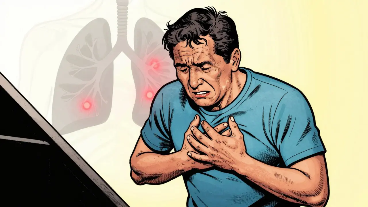
You think the worst part is over. You tested negative, you survived the acute phase, and yet, you still can’t catch your breath climbing stairs. It feels like there is something wrong with your engine, but standard tests come back saying everything is fine. Pulmonary Long COVID is a persistent respiratory condition characterized by lingering lung function issues after viral clearance. It turns out, the virus left traces in your smallest airways that regular scans miss.
Recent research from 2025 confirms that this isn’t just "feeling tired." There is actual physiological damage occurring deep within the lung tissue. About one-third of all long-term post-viral cases involve specific lung complications. If you are struggling months later, you aren’t imagining it. Understanding the mechanism helps you know which doctors to see and what treatments actually work.
What Actually Happens Inside Your Lungs
To understand why you feel short of breath, we need to look at the immune system’s overreaction. When your body fought off SARS-CoV-2 is the coronavirus strain responsible for the global pandemic and subsequent long-term symptoms., it deployed neutrophils. These are white blood cells designed to eat germs. But here’s the problem: sometimes they don’t stop fighting when the enemy is gone.
In late 2023 through 2025, researchers led by Dr. Don Sin at the Centre for Heart Lung Innovation found that these neutrophils keep triggering an immune response even after the virus clears. They call them “dirty bombs.” They continue damaging the small airways where oxygen transfer happens. Standard CT scans often can’t see this damage because the large airways look normal, but the tiny gas-exchange units are inflamed. This leads to a mismatch where your lungs can’t efficiently get oxygen into your bloodstream, resulting in fatigue and breathlessness during physical exertion.
Advanced Diagnostics Beyond Standard Tests
If your doctor says your lung function tests are “within range,” does that mean you are healed? Not necessarily. Traditional spirometry measures airflow volume, like FEV1 is Forced Expiratory Volume in one second, a key metric in assessing lung capacity. It tells you how fast you blow air out, but it doesn’t show how well that air exchanges oxygen. That’s where newer technology comes in.
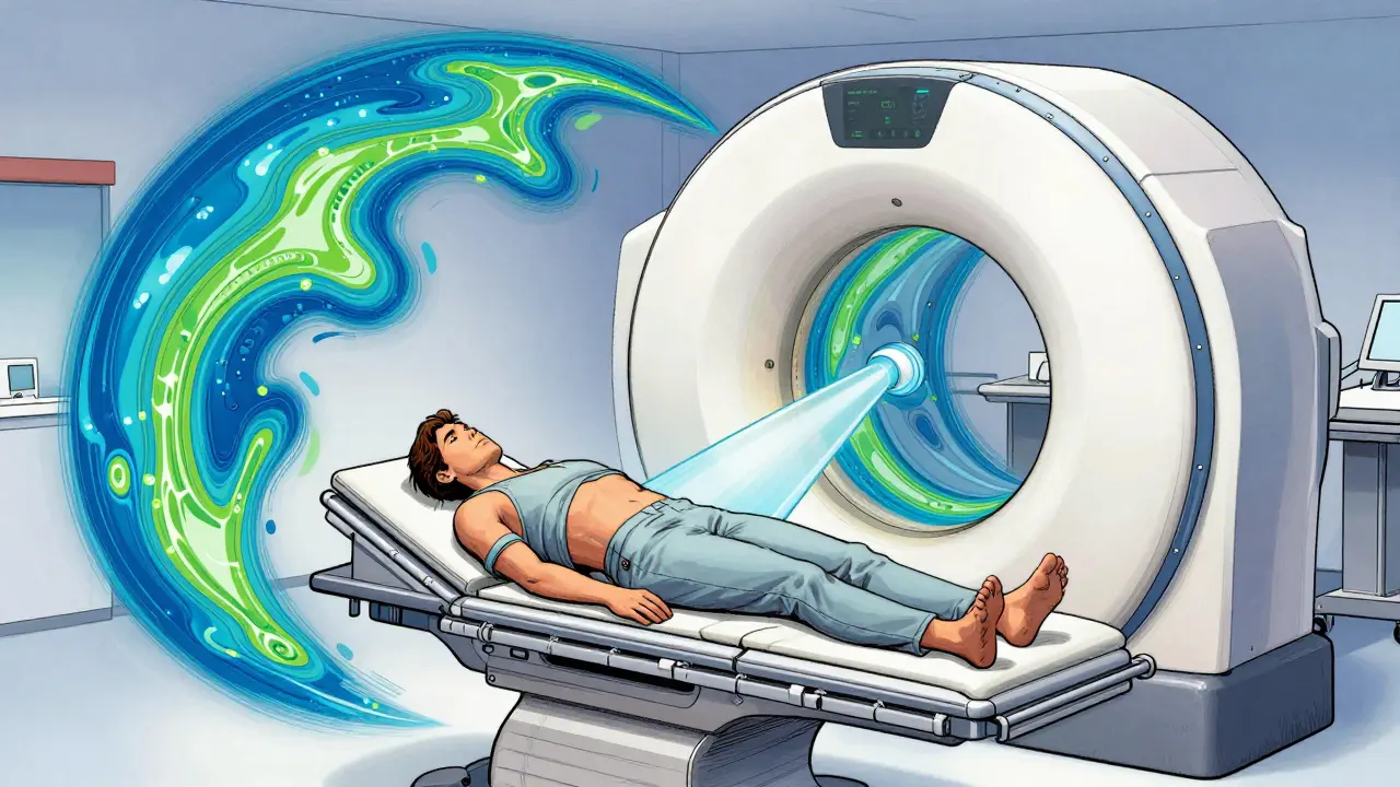
Hyperpolarized Xenon MRI is an advanced imaging technique using inert gas to visualize gas exchange in small airways. Think of it as a specialized video camera for the deepest parts of your lungs. A team at the University of British Columbia successfully used this in 2024 and 2025 to map exactly where the inflammation is hiding. They identified four distinct clusters of damage. For patients who don’t improve with standard care, this scan provides the roadmap for targeted therapy. While not yet available in every clinic, major centers like Duke and the University of Kansas are adopting it.
| Test Method | What It Measures | Limitations |
|---|---|---|
| Standard Spirometry | Airflow volume (FEV1) | Misses small airway damage |
| Xenon MRI | Oxygen transfer efficiency | Requires specialized equipment |
| CT Scan | Structural changes/Fibrosis | Radiation exposure, misses subtle inflammation |
Risk Factors: Who Needs Extra Watch?
Not everyone walks this path equally. Data from a South Korean study covering 688 patients showed significant differences based on severity. Among those hospitalized for severe infection, roughly 12.6% developed Post-COVID-19 Pulmonary Fibrosis is permanent scarring of lung tissue that reduces elasticity and gas exchange.. Scarring means your lungs become stiff like dry leaves instead of soft balloons.
If you had underlying conditions before the infection, your starting point was lower. People with Chronic Obstructive Pulmonary Disease is a group of chronic inflammatory lung diseases causing obstructed airflow. faced higher risks. The stats were stark: those with COPD who caught COVID had a mortality rate of 4.6%, compared to zero percent for non-COVID patients in some cohorts. They also suffered more acute flare-ups afterward. If you fall into this category, monitoring becomes critical because your margin for error is smaller.
Hospitalization status matters less than you might think regarding breathing issues later on. One Australian review found that mechanical ventilation didn’t predict worse lung function three months out compared to less intensive care. However, being admitted did correlate with a 2.60-fold higher risk of persistent breathlessness. It suggests the intensity of the initial infection drives the long-term damage rather than just the machines used to treat it.
Rehabilitation: Moving Toward Function
Waiting for your body to heal on its own isn’t usually enough. The muscles around your chest forget how to move correctly, and your cardiovascular system deconditions quickly. A structured program is essential. Pulmonary Rehabilitation is a comprehensive intervention including exercise training and education tailored to lung health. typically starts four weeks after the acute phase ends.
Effective programs run for eight to twelve weeks. Sessions happen two or three times a week. The mix is important: you need breathing exercises to retrain diaphragm control, aerobic conditioning to rebuild stamina, and strength training to support movement. A pre-and-post study showed participants improved their diffusion capacity and walked further on the six-minute walk test after completing the course.
It isn’t just about gym time; it’s about pacing. Many patients experience Post-Exertional Malaise is a worsening of symptoms following physical or mental exertion. pushing too hard makes things worse. Clinicians use the 30-second sit-to-stand test (30STS) to gauge progress safely. The goal is gradual improvement without triggering a crash.

Tracking Your Progress
How do you know if it’s working? Subjective feelings matter, but objective metrics help. Doctors use the mMRC dyspnea scale to grade breathlessness. A score of two or higher at the one-month mark often predicts residual dysfunction. This gives early warning signs that you need more aggressive rehab.
Look for trends over six months. Most moderate to severe cases show measurable improvement in lung function over that period. However, the curve flattens eventually. If you aren’t seeing gains after three months of consistent effort, it’s time to request advanced diagnostics like the Xenon MRI mentioned earlier. Don’t settle for a diagnosis of “it’s just anxiety” when your lungs have been injured by inflammation.
Therapeutic Considerations
Treatment isn’t one-size-fits-all. The drug you took during the infection might have influenced your outcome. Remdesivir use appeared linked to a lower risk of fibrosis in some analyses, while Baricitinib showed increased risk, though researchers warn these links need more validation. Future therapies aim to target the neutrophil activity directly. Clinical trials planned for 2026 focus on stopping that chronic low-grade inflammation. Until then, managing symptoms and rebuilding capacity remain the priorities.
Frequently Asked Questions
Can lung damage from COVID be reversed?
Some inflammation resolves with time and rehabilitation, but permanent scarring known as fibrosis may remain. Research shows functional improvements are possible even with structural changes.
What test finds hidden lung damage?
Hyperpolarized Xenon MRI is superior for visualizing small airway gas exchange abnormalities that standard CT scans often miss.
How long should rehabilitation last?
Standard protocols last 8 to 12 weeks, typically starting 4 weeks post-infection, with sessions scheduled 2-3 times weekly.
Does prior COPD increase long-term risk?
Yes, patients with pre-existing COPD face significantly higher mortality rates and more frequent exacerbations following infection compared to those without.
When will symptoms start improving?
Most measurable improvements in lung function occur within the first six months, though some residual effects may persist for years.